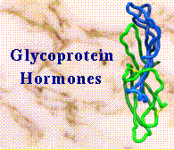

The glycoprotein hormones are a family of cystine rich proteins consisting of an alpha and beta subunit. The alpha subunit is common to each of the hormones for a given species. The beta subunit is assumed to confer the hormone specificity. The glycoprotein hormones are characterised by there heavy glycosylation and are cystine rich molecules with 5 conserved disulphides in alpha and 6 in beta.
Here are a quick access to glycoprotein hormone sequences.
 Alpha Subunit
  Chorionic gonadotropin Beta Subunit
 Luteinizing hormone Beta Subunit
 Follicle Stimulating Beta Subunit
 Thyroid Stimulating Beta Subunit
The prosite sequence signatures.
 Alpha Subunit
 Beta Subunit
The structure of hCG was first determined in Glasgow at 3.0Ang resolution and is described in the paper 'Crystal Structure of Chorionic Gonadotropin'. [Lapthorn AJ, Harris DC, Littlejohn A, Lustbader JW, Canfield RE, Machin KJ, Morgan FJ & Isaacs NWI. Nature 369, 455-461 (1994).] This was followed closely by a 2.6Ang resolution structure from Wayne Hendrickson's group. [Wu H, Lustbader JW, Liu Y, Canfield RE, Hendrickson WA. Structure 2, 545-558 (1994).]
Various graphical representations of the 3D structure of hCG. This includes a Hyperactive Molecule of hCG inspired by those clever chaps at imperial.
The crystal structure of hCG has allowed the correct assignment of the disulphide linkages of both the alpha and beta subunits. The assignments determined chemically by Mise and Bahl which were generally accepted as the most reliable were shown to be partially correct for both subunits.
With the correct disulphide assignments it became apparent that both the alpha and beta subunits had an unusual "knot" of cystines. This unusual feature has been seen in a number of protein growth factors namely Nerve growth factor, Platelet derived growth factor BB and Transforming growth factor B. The cystine knot comprises three disulphide bridges, arranged so that two disulphides link adjacent parallel strands of the peptide chain and form a ring through which the third disulphide penetrates.
The structure of each subunit is essentially the same .Each molecule has two beta-hairpin loops on one side of a central cystine knot and a long loop on the other. In the beta-subunit the beta-hairpins are stabilized by the disulphide 23-72.
Superimposing the alpha and beta-subunits at their cystine-knot shows clearly the common fold and the extent of their common structure. The CA positions for 22 residues extending from the knot can be superimposed with a r.m.s. difference of 0.52A. The beta-subunit clearly has extended beta-hairpins and a longer C-terminus than the alpha-subunit.