Compact myelin of the central nervous system (CNS) contains a high proportion of lipid (ca. 70 %) and several proteins, which are highly enriched in, or specific to, myelin. The highly hydrophobic transmembrane proteolipid protein, (PLP), and the extrinsic, hydrophilic myelin basic protein (MBP), are the major myelin proteins of the CNS accounting for approximately 40 % and 30 %, respectively of total protein. Although the myelin proteins are largely conserved across species, our initial investigations have shown that while similar to other well-defined myelin proteins ( e.g. bovine, human, rat, murine) equine myelin has some distinct differences. This work is primarily concerned with PLP, which is one of the most hydrophobic proteins in nature, and its DM20 isoform, which differs from PLP by the insertion of an additional 35 amino acid cytoplasmic domain near the centre of the protein. In PLP, the 276 amino acid polypeptide are thought to be structured into four hydrophobic, membrane spanning, and five hydrophilic domains (see below). PLP and DM20 are both involved in the formation of the intraperiod line (IPL), which is essential to the correct architecture of the myelin sheath. Both proteins are implicated in axon/glial interactions and patients lacking PLP and/or DM20 develop axonopathy. Mutations of the X-linked PLP gene cause the neurological disorders Pelizaeus-Merzbacher disease (PMD) and Spastic Paraplegia type 2 (SPG2) and similar disorders in a variety of animals . A current conundrum is how to link the molecular defects with the heterogeneous phenotypes. This task would be simplified if we understood the 3-dimensional structures that PLP and DM20 adopt in the oligodendrocyte membrane.
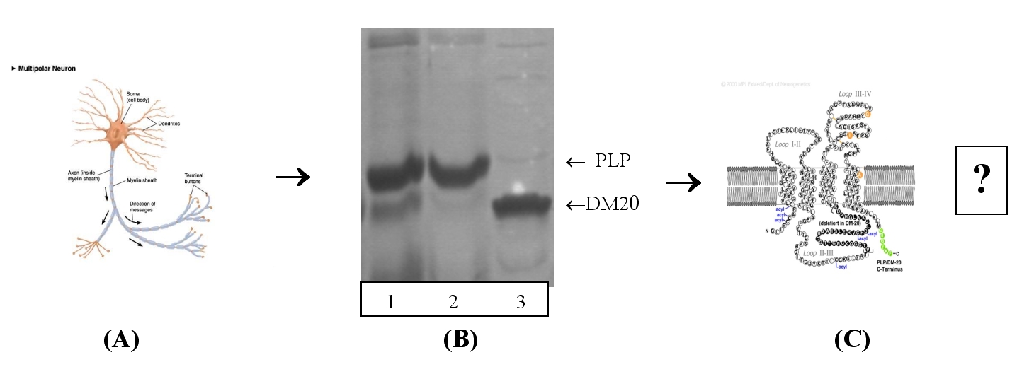
Figure shows : (A) Nerve cell with the axon coated in myelin sheath. PLP and its DM20 isoform. To date most of our work is focussed on the extraction and purification of these transmembrane proteins using specific detergents to solubilize the lipid bilayer without compromising the structural integrity of the proteins. Interestingly, we routinely extract PLP and DM20 as individual components and also as a complex. Currently, we are conducting crystallization trials on all three entities. (Shown above on SDS-PAGE, 1: PLP/DM20 complex, 2: PLP and 3: DM20) We are trying to correlate the protein profiles which are obtained from detergent solubilised samples obtained from transgenic mice with and without mutations collected over different life time spans. Relevant publications: Dominic J.B. Hunter, Rachel Macmaster, Aleksander W. Roszak, Alan Rimbaldi-Tunnicliffe, Ian R. Griffiths and A. Freer . (2005) Structure of myelin P2 protein from Equine Spinal Cord. Acta Cryst D61 , pp1067-1071. McLaughlin M., Hunter D., Thompson C., Yool D., Kirkham D., Freer A. & Griffiths I.(2002) Evidence for possible interactions between PLP and DM20 within the myelin sheath. Glia, 39 , Issue 1, pp 31-36. Wood D, She Y, Freer A , Harauz G. & Moscarello A. (2002) Primary structure of equine myelin basic protein by mass spectrometry. Arch. Biochem. & Biophys . 405 , pp 137-146.
|