Nanomaterials Photo Gallery
Nanomaterials have many potential technological applications, but in addition, images of nanomaterials are often stunning.
Click here for further information (a link to the Materials and Condensed Matter Physics research group webpages)
Below, you'll find a selection of images of some of the nanomaterials made recently in our lab, each with an explanatory note.
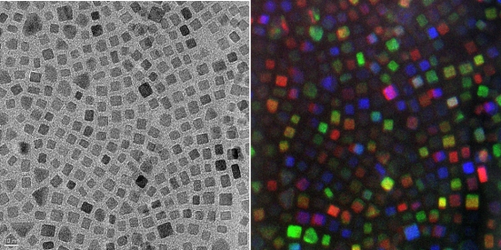 |
Bright field and corresponding false-coloured dark field TEM image of an assembly of cubic iron oxide nanoparticles (picture taken by F. Douglas using the T20 microscope in the School of Physics in collaboration with Dr D. MacLaren) |
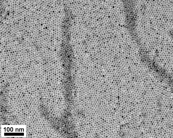 |
TEM image of an iron oxide nanoparticle superlattice. Superlattice formation occurs when there is a narrow distribution of particle sizes (picture taken by F. Douglas using the T20 microscope in the School of Physics in collaboration with Dr D. MacLaren)
|
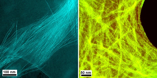 |
False-coloured dark field images of lanthanide oxide nanowires (picture taken by F. Douglas using the TF20 microscope in the School of Physics in collaboration with Dr D. MacLaren) |
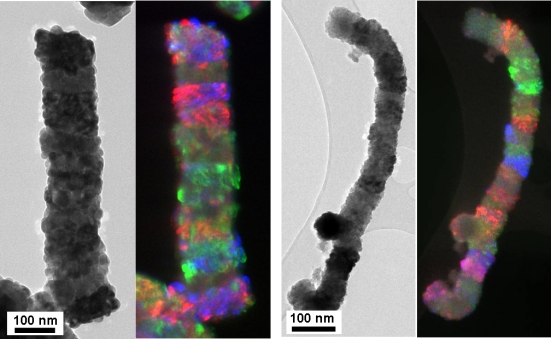 |
Bright field and corresponding false-coloured dark field images of two iron oxide nanospecies. Dark field imaging was used to show that these species grow by oriented assembly of smaller, cubic subunits (picture taken by F. Douglas using the T20 microscope in the School of Physics in collaboration with Dr D. MacLaren) |